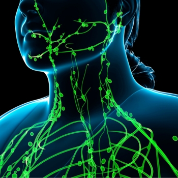
Detoxification is essential for maintaining health. It is the body’s natural process of identifying, breaking down, and eliminating potentially harmful substances. The liver does most of this work, while the kidneys, digestive system, lungs, skin, and lymphatic system help remove waste through urine, stool, breath, and sweat. The body continuously detoxifies itself; it does not require special cleanses or extreme diets to perform these essential functions. Several mechanisms in the body accomplish this work.
Two of the most important are the glymphatic and lymphatic systems.
The Glymphatic System
The glymphatic system is the brain’s waste-clearance pathway. It relies on glial cells and cerebrospinal fluid to remove metabolic waste, especially during deep sleep.
Two types of fluid work together to move waste:
- Cerebrospinal fluid (CSF): CSF surrounds and protects your brain and spinal cord. It enters the brain through small spaces surrounding blood vessels, known as perivascular or Virchow-Robin spaces. The fluid moves as the blood vessels expand and contract, creating small waves with your heartbeat and breathing.
- Interstitial fluid (ISF): ISF surrounds and protects tissues and cells. CSF meets ISF as it travels through brain tissue, aided by aquaporin-4 (AQP4) water channels. AQP4 is a protein that helps move water through your organs. Waste collection occurs naturally as CSF and ISF move through brain tissue.
ISF carries the collected waste out of the brain through perivascular spaces. The waste then enters the lymphatic system in the neck.
How It Works
- Fluid flow: Cerebrospinal fluid flows into the brain along arteries, moves through tissue, and mixes with the fluid between brain cells.
- Cell channels: Aquaporin-4 water-channel proteins on brain cells called astrocytes help move the fluid.
- Waste removal: The fluid collects unwanted waste and drains along veins toward the neck. The brain needs fuel to work, and using that fuel leaves metabolic waste that must be removed.
Why Is It Critical for Brain Health?
In Alzheimer’s disease, for example, major changes in the brain include the accumulation of amyloid-beta and tau proteins. The brain must also manage metabolic byproducts such as lactic acid and excess potassium. The glymphatic system helps clear excess amyloid-beta, tau, and other waste.
The glymphatic system also circulates essential sugars, lipids, amino acids, and neurotransmitters needed for communication between cells.
The system works most actively during sleep, when changes in the space between brain cells allow fluid to flow more easily.
Health Conditions That Negatively Affect the Glymphatic System
A growing body of research links impaired glymphatic function with several neurological and psychiatric conditions. Sleep disorders are especially important because much of the system’s waste-removal activity occurs during sleep. Inadequate or disrupted sleep may interfere with the movement of cerebrospinal fluid and the clearance of metabolic waste from brain tissue. Researchers have also reported possible connections among glymphatic dysfunction, migraine and other headaches, depression, anxiety, and additional mood disorders. However, evidence concerning headaches and mood disorders is still developing, and researchers have not established whether altered glymphatic function is a cause, consequence, or contributing factor. 1 2
Conditions that injure, inflame, or cause abnormal protein accumulation in the brain may also interfere with glymphatic circulation. Stroke, traumatic brain injury, and meningitis can alter fluid movement, damage blood vessels, or produce swelling that obstructs normal waste clearance. Glymphatic impairment has also been observed in neurodegenerative disorders, including Alzheimer’s disease and Parkinson’s disease. In these disorders, reduced clearance may contribute to the accumulation of proteins such as amyloid-beta, tau, and alpha-synuclein. Scientists are still investigating whether glymphatic dysfunction helps initiate these diseases or develops as the brain becomes damaged. 3 4
How to Support the Glymphatic System
- Get a good night’s sleep. Go to bed at the same time each night and maintain a regular sleep schedule. Develop a relaxing bedtime routine. Turn off televisions, phones, and computers at least one hour before bedtime. Avoid heavy meals or alcohol before sleeping, and have your last meal or snack at least two hours before bedtime.
- Exercise regularly.
- Stay hydrated. Drink plenty of fluids.
- Eat well-balanced meals.
- Manage stress.
The Lymphatic System
The lymphatic, or lymphoid, system is part of the circulatory system. It consists of a vital network of vessels, nodes, and organs, including the lymph nodes, spleen, and thymus. It helps maintain fluid balance, fight germs, and absorb fats. It acts as the body’s cleanup and defense system by capturing bacteria, viruses, and abnormal cells and helping destroy them.
Lymph is a clear fluid that moves from the blood into tissues, where it collects waste, bacteria, and white blood cells. Lymph nodes are small, bean-shaped glands that filter this fluid and trap germs and damaged cells.
Lymphatic vessels are tiny tubes that collect lymph and carry it in one direction back toward the heart.
The spleen and thymus are particularly important organs. They filter blood and help produce or support specialized white blood cells called lymphocytes, which fight infection.
The lymphatic system also absorbs dietary fats from the digestive tract and moves them into the bloodstream.
The central nervous system was long considered to be devoid of lymphatic vessels until researchers identified them in the cranial meninges. 5 6 Moreover, researchers have observed a vessel with lymphatic features in the eye. 7 8
How to Support the Lymphatic System
You can support your lymphatic system through regular physical activity, deep breathing, and proper hydration. Unlike blood, lymph fluid has no central pump like the heart. It relies on body movement, muscle contractions, and pressure changes to circulate.
- Take daily walks: The calf muscles act as a natural pump for fluid circulation.
- Use dry brushing: Brush dry skin with long, light strokes toward your heart before showering to stimulate surface circulation.
- Practice belly breathing: Deep diaphragmatic breathing creates pressure changes in the chest that help move lymph fluid through major pathways.
- Eat clean foods: Focus on fresh fruits, vegetables, and lean proteins while cutting back on highly processed foods such as chips, cheese puffs, packaged snack cakes, hot dogs, and bacon.
- Drink plenty of water: Proper hydration supports normal lymph flow.
Microglia Eliminate Brain Waste
Microglia are the resident macrophages and primary immune cells of the brain. They perform many functions, including attacking and consuming bacteria through phagocytosis, clearing cellular waste, providing neuroprotection, and supporting the growth of new neurons. They interact with astrocytes, neurons, and endothelial cells.
Microglial cells help break down unnecessary or dead nerve tissue. 9 10 These cells are part of the immune system. In conditions such as Alzheimer’s disease, they may fail to clear waste effectively, including beta-amyloid deposits. 11
How Do the Eyes Eliminate Waste?
Waste elimination in the eyes is critical for supporting and maintaining healthy vision. Many leading causes of irreversible blindness, including glaucoma and age-related macular degeneration, involve the buildup of fluid, waste, or inflammatory debris in or near the back of the eye.
The retina is one of the most metabolically active tissues in the body. It constantly generates byproducts that must be cleared. In dry macular degeneration, for example, some waste is not fully reabsorbed and eliminated. Deposits called drusen can accumulate beneath the retina. Over time, these deposits are associated with damage to the underlying retinal cells and declining vision.
The eyes eliminate waste through specialized cellular recycling, fluid-drainage systems, and tear flushing. Retinal pigment epithelial cells break down intracellular debris via autophagy and phagocytosis. Fluid and metabolic waste drain through internal pathways such as the trabecular meshwork and the recently studied posterior ocular lymphatic outflow pathway. Tears wash away surface debris and dead cells.
Nutrients That Help the Eyes
The good news is that targeted nutrients may help strengthen and protect the retina while supporting its ability to manage oxidative stress and metabolic waste. These nutrients do not physically “clean” the retina. Instead, they may protect retinal cells, improve circulation, filter damaging light, and support antioxidant systems that neutralize or remove harmful byproducts.
- Taurine: The retina contains one of the highest concentrations of taurine in the body. This amino acid helps stabilize retinal cell membranes, regulate calcium, and protect photoreceptors and retinal ganglion cells from oxidative stress. Taurine deficiency has been associated with retinal degeneration, suggesting that adequate levels are essential for retinal health. 12
- Lutein: Lutein accumulates in the macula, where it helps form the protective macular pigment. It filters some high-energy blue light and helps neutralize free radicals before they damage retinal tissue. Supplementation may increase macular pigment density and improve retinal sensitivity in people with early age-related macular degeneration. 13
- Zeaxanthin: Zeaxanthin is concentrated near the center of the macula, the area responsible for sharp, detailed vision. Along with lutein, it filters blue light and helps protect retinal cell membranes from oxidation. Clinical research indicates that lutein and zeaxanthin supplementation can increase macular pigment and may improve visual function in people with early macular degeneration. 14
- Meso-zeaxanthin: Meso-zeaxanthin is found primarily in the central macula, where visual detail is the sharpest. Together, lutein, zeaxanthin, and meso-zeaxanthin create a protective pigment that absorbs blue light and helps limit oxidative damage. Supplementation with all three carotenoids may produce a more complete increase in macular pigment than formulas that do not contain meso-zeaxanthin. 15
- Astaxanthin: Astaxanthin is a red carotenoid with antioxidant and anti-inflammatory properties. It may help protect retinal cells and their mitochondria, which produce the energy needed for vision but also generate oxidative waste. In one clinical trial, a supplement containing astaxanthin and several other antioxidants improved measurements of central retinal function in people with nonadvanced macular degeneration. 16
- Ginkgo biloba: Ginkgo contains antioxidant plant compounds and may support blood flow through the tiny vessels that supply the retina and optic nerve. Healthy circulation helps deliver oxygen and nutrients while carrying away carbon dioxide and metabolic byproducts. A small controlled trial found improved visual-field measurements in some patients with normal-tension glaucoma, although larger studies are needed. 17
- Bilberry: Bilberry contains anthocyanins, pigments with antioxidant and blood-vessel-supporting properties. These compounds may help protect delicate eye tissues from oxidative stress and support microcirculation. In a human trial, bilberry supplementation reduced several symptoms of screen-related eye fatigue, although research on its ability to prevent retinal disease remains limited. 18
For more information on visual detox, visit Visual Detox: A Whole-Body Connection.
Foods That Support the Body’s Natural Detoxification Processes
Food can provide fiber, antioxidants, sulfur compounds, and other nutrients involved in the body’s detoxification and waste-removal systems. Several types of food stand out.
- Garlic and onions: Cuisines around the world use these two root vegetables heavily. Eastern European countries, for example, add garlic and onions to many daily meals. In India, they are valued as medicines. Sulfur-containing compounds in these vegetables may support glutathione production and the enzymes involved in processing certain substances. Human research exists, although much of the evidence about the mechanisms comes from laboratory and animal studies. Garlic supplementation has also been associated with improved antioxidant markers. 19
- Cruciferous vegetables: Broccoli, kale, Brussels sprouts, and cabbage contain glucosinolates. These substances produce compounds such as sulforaphane, which affect enzymes the body uses to process and eliminate foreign chemicals. A human trial found that a broccoli-sprout beverage increased the urinary elimination of certain airborne pollutants. 20
- Carrots, seeds, nuts, and vegetables: Their fiber supports regular bowel movements and helps the body eliminate waste through the digestive tract.
Cucumber Detox Recipe
This low-calorie drink supports daily hydration, aids digestion, and provides small amounts of vitamins and antioxidants without added sugar.
Ingredients
- 1 English cucumber
- 1 lemon
- Fresh mint
- 2 quarts (8 cups) drinking water
- Slice the cucumber and lemon.
- Add the slices to a large pitcher with a few sprigs of fresh mint.
- Pour in the water.
- Place the pitcher in the refrigerator for at least two hours to allow the flavors to blend.
Suggested Supplements to Consider
- Dr. Grossman’s Complete Eye Formula 2 oz (oral spray)
- ACG Glutathione EXTRA STRENGTH Spray 2 oz
- Dr. Grossman’s Meso Plus Formula with Astaxanthin – 90 vcaps (3-month supply)
- Advanced Eye & Vision Support Formula (whole food) 60 vcaps
- ReVision Formula (wild-crafted herbal formula) 2 oz – Based on a classic Chinese medicine liver tonic formula to help support healthy circulation and blood flow throughout the eyes and body. It also contains milk thistle extract to support liver detoxification.
- Dr. Grossman’s Retinal Support (wild-crafted herbal formula) 2 oz – Helps support the kidney and spleen meridians, which are related to healthy blood support, digestion and distribution of food and nutrients, and bone, back, and knee support.
- Visual Detox Homeopathic Drops 2 oz
- Dr. Grossman’s Blood Vessel Control Formula 2 oz – Helps reduce the risk of unwanted blood-vessel growth, supports healthy circulation, and reduces inflammation.
- Dr. Grossman’s Bilberry/Ginkgo Combination 2 oz (60 ml) – Helps support healthy circulation and strengthen blood vessels and capillaries.
- Dr. Grossman’s Whole Food Organic Superfood Multi-Vitamin 120 vcaps – Whole-food, organic, GMO-free multivitamin.
- H2 Elite Molecular Hydrogen 60 tabs
- NMN Wonderfeel Capsules 60 vegcaps – Helps protect retinal ganglion cells and optic nerves from damage, reduce inflammation, and support the immune system.
Supplement Packages
- Dr. Grossman’s Complete Eye (oral spray)/Meso Plus Combo Package
- Ultimate Body Detox Extra Strength 2 oz Bottles – This unparalleled detoxification program supports immune function, detoxification, and rejuvenation, featuring three powerful ingredients. The Ultimate Body Detox regimen quickly and effectively reduces Toxic Body Burden (TBB), defined as the cumulative and damaging relationship among pathogens, toxins, and free radicals. The destructive sum is greater than the whole. To reduce TBB, pathogens and toxins must be eliminated and free radicals neutralized through an effective, multifaceted approach that provides both treatment and prevention.
- AMD Package 1 (3-month supply)
- Brain and Memory Support Package 1
- Anti-Inflammatory Eye and Whole Body Package 1 (1-month supply)
Recommended Books
- Natural Eye Care: Your Guide to Healthy Vision and Healing
- Natural Brain Support: Your Guide to Preventing and Treating Alzheimer’s, Dementia, and Other Related Diseases Naturally
- Natural Parkinson’s Support: Your Guide to Preventing and Managing Parkinson’s
- Fretes Burgos A, Olson PA, Vgontzas A. “The Glymphatic System and Its Relationship to Migraine.” Current Pain and Headache Reports. 2024;28(9):867-875. https://pubmed.ncbi.nlm.nih.gov/39150650/ ↩
- De Berardis D, et al. “Glymphatic System and Psychiatric Disorders: A Rapid Comprehensive Scoping Review.” Current Neuropharmacology. 2025;23(3):275-287. https://pubmed.ncbi.nlm.nih.gov/39234773/ ↩
- Lee DA, et al. “Magnetic Resonance Images Implicate That Glymphatic Alterations Mediate Cognitive Dysfunction in Alzheimer Disease.” Annals of Neurology. 2022;93(1):164-174. https://pubmed.ncbi.nlm.nih.gov/36214568/ ↩
- Ma X, et al. “Diffusion Along Perivascular Spaces as Marker for Impairment of Glymphatic System in Parkinson’s Disease.” NPJ Parkinson’s Disease. 2023;9(1):13. https://pubmed.ncbi.nlm.nih.gov/36543809/ ↩
- Louveau A, Smirnov I, Keyes TJ, et al. “Structural and Functional Features of Central Nervous System Lymphatic Vessels.” Nature. 2015;523(7560):337-341. ↩
- Aspelund A, Antila S, Proulx ST, et al. “A Dural Lymphatic Vascular System That Drains Brain Interstitial Fluid and Macromolecules.” Journal of Experimental Medicine. 2015;212(7):991-999. ↩
- Kizhatil K, Ryan M, Marchant JK, Henrich S, John SW. “Schlemm’s Canal Is a Unique Vessel With a Combination of Blood Vascular and Lymphatic Phenotypes That Forms by a Novel Developmental Process.” PLoS Biology. 2014;12(7):e1001912. ↩
- Aspelund A, Tammela T, Antila S, et al. “The Schlemm’s Canal Is a VEGF-C/VEGFR-3-Responsive Lymphatic-Like Vessel.” Journal of Clinical Investigation. 2014;124(9):3975-3986. ↩
- Bessis A, Bechade C, Bernard D, Roumier A. “Microglial Control of Neuronal Death and Synaptic Properties.” Glia. 2007;55(3):233-238. ↩
- Caldero J, Brunet N, Ciutat D, Hereu M, Esquerda JE. “Development of Microglia in the Chick Embryo Spinal Cord: Implications in the Regulation of Motoneuronal Survival and Death.” Journal of Neuroscience Research. 2009;87(11):2447-2466. ↩
- National Institute on Aging. “What Happens to the Brain in Alzheimer’s Disease?” https://www.nia.nih.gov/health/alzheimers-causes-and-risk-factors/what-happens-brain-alzheimers-disease. ↩
- Castelli V, et al. “Taurine and Oxidative Stress in Retinal Health and Disease.” CNS Neuroscience & Therapeutics. 2021;27(4):403-412. https://pubmed.ncbi.nlm.nih.gov/33621439/ ↩
- Huang YM, et al. “The Effects of Lutein and Zeaxanthin Supplementation on Visual Performance in Patients With Age-Related Macular Degeneration.” British Journal of Nutrition. 2015;114(3):406-412. https://pubmed.ncbi.nlm.nih.gov/25228440/ ↩
- Ma L, et al. “Effect of Lutein and Zeaxanthin on Macular Pigment and Visual Function in Patients With Early Age-Related Macular Degeneration.” Ophthalmology. 2012;119(11):2290-2297. https://pubmed.ncbi.nlm.nih.gov/22858124/ ↩
- Loughman J, et al. “Macular Pigment Response to Lutein, Zeaxanthin, and Meso-zeaxanthin Supplementation in Open-Angle Glaucoma.” Ophthalmology Science. 2021;1(4):100039. https://pubmed.ncbi.nlm.nih.gov/36247822/ ↩
- Parisi V, et al. “Carotenoids and Antioxidants in Age-Related Maculopathy Italian Study: Multifocal Electroretinogram Modifications After 1 Year.” Ophthalmology. 2008;115(2):324-333.e2. https://pubmed.ncbi.nlm.nih.gov/17716735/ ↩
- Quaranta L, et al. “Effect of Ginkgo biloba Extract on Preexisting Visual Field Damage in Normal Tension Glaucoma.” Ophthalmology. 2003;110(2):359-364. https://pubmed.ncbi.nlm.nih.gov/12578781/ ↩
- Ozawa Y, et al. “Bilberry Extract Supplementation for Preventing Eye Fatigue in Video Display Terminal Workers.” Journal of Nutrition, Health & Aging. 2015;19(5):548-554. https://pubmed.ncbi.nlm.nih.gov/25923485/ ↩
- Askari M, Mozaffari H, Darooghegi Mofrad M, et al. “Effects of Garlic Supplementation on Oxidative Stress and Antioxidative Capacity Biomarkers: A Systematic Review and Meta-Analysis of Randomized Controlled Trials.” Phytotherapy Research. 2021;35(6):3032-3045. https://pubmed.ncbi.nlm.nih.gov/33484037/ ↩
- Egner PA, Chen JG, Zarth AT, et al. “Rapid and Sustainable Detoxication of Airborne Pollutants by Broccoli Sprout Beverage: Results of a Randomized Clinical Trial in China.” Cancer Prevention Research. 2014;7(8):813-823. https://pubmed.ncbi.nlm.nih.gov/24913818/ ↩