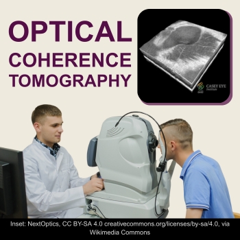
The fact that the retina and optic nerve are brain tissue could give doctors instant insight into Alzheimer’s disease. Modern eye-scanning technology is beginning to detect brain problems by examining the retina. Non-invasive exams and OCT (Optical Coherence Tomography) scans can detect the early appearance of beta-amyloid plaques and tau proteins. Plaques and tau proteins are found in the brains of patients with Alzheimer’s disease. Their appearance in the retina may indicate early signs of Alzheimer’s disease. The earlier Alzheimer’s is caught, the more time a patient has to head it off, particularly through diet, exercise, and targeted supplementation.
Unlike the brain, the retina can be photographed and measured quickly, without surgery or ionizing radiation. A scientific review published in July 2026 examines the growing evidence that Alzheimer’s-related changes may affect retinal nerve cells, blood vessels, supporting cells, and proteins. 1 The findings are encouraging, but they do not mean that an eye examination can currently tell you whether you have Alzheimer’s disease.
Why Alzheimer’s Disease May Affect Your Eyes
The retina is part of your central nervous system. It develops from the same embryonic tissue as the brain and contains neurons, immune cells, blood vessels, and protective barriers that are similar to those in the brain. 2
Alzheimer’s disease involves much more than memory loss. Biological changes may begin many years before noticeable symptoms appear. These changes include the accumulation of amyloid-beta, abnormal forms of the protein tau, inflammation, impaired circulation, and the gradual loss of connections between nerve cells.
Some of these processes may also occur in the retina. In addition, Alzheimer’s disease can affect the parts of the brain that interpret visual information. This means that a person may have difficulty recognizing objects, judging depth, reading, detecting contrast, or navigating a room even when the eyes themselves remain capable of seeing.
Alzheimer’s-Related Proteins in the Retina
One of the most closely studied questions is whether amyloid-beta can accumulate in the human retina.
In a 2017 study, researchers examined retinas from people with confirmed Alzheimer’s disease and reported amyloid deposits, particularly in the upper and lower peripheral retina. The investigators also conducted a small proof-of-concept trial using curcumin, a naturally fluorescent plant compound, to help make possible retinal amyloid deposits visible in living patients. 3
Other laboratories have not always found amyloid-beta in the same amounts or at the same locations. Different tissue-processing methods, antibodies, staining techniques, disease stages, and definitions of a positive deposit can affect the results. One postmortem study found amyloid-beta and phosphorylated tau in some retinas but concluded that the retinal changes did not clearly separate Alzheimer’s cases from controls. 4
Researchers have also detected abnormal tau, activated immune cells, and signs of inflammation in the retina. 5 A subsequent human tissue study found that the amount and distribution of phosphorylated tau in the retina were associated with tau pathology in the brain. 6
In 2023, a large study combined retinal tissue examination with protein analysis. The researchers reported that the retinal tissue contained amyloid-beta plaques, blood-vessel abnormalities, evidence of inflammation, and changes in proteins involved in energy production and cell survival. Several retinal findings correlated with cognitive scores and the severity of brain disease. 7
These findings strengthen the case that the retina participates in Alzheimer’s-related disease processes. However, tissue examined after death is very different from retinal tissue scanned during an office visit.
Changes in Retinal Nerve Tissue
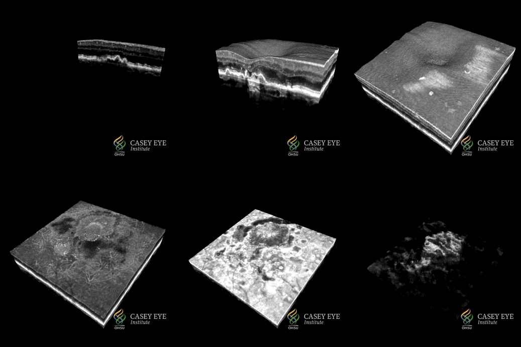
Optical coherence tomography, usually called OCT, uses reflected light to produce detailed cross-sectional images of the retina. It can measure individual retinal layers to within a few thousandths of a millimeter. Eye doctors already use OCT to monitor glaucoma, macular degeneration, diabetic eye disease, and other conditions.
Many studies have found that people with Alzheimer’s disease have thinning of the retinal nerve fiber layer and ganglion cell layer. These layers contain nerve cells and fibers that help carry visual information from the eye toward the brain.
A 2017 systematic review found significant thinning in several retinal regions among people with Alzheimer’s disease. 8 A larger 2019 analysis also found reduced thickness in multiple retinal layers, although the results differed according to disease severity, retinal location, and study methods. 9
Thinning is not the only reported pattern. Some studies of people in the earliest stages have found temporary thickening in certain layers. Inflammation or an early response to injury may occur before tissue is lost. This possibility is still being investigated.
What Tiny Blood Vessels May Reveal
The retina contains a network of blood vessels that can be viewed directly. Retinal photography shows the larger vessels, while OCT angiography, or OCTA, maps blood flow through very small vessels without requiring an injected dye.
Researchers have reported narrower vessels, less complex branching, lower vessel density, reduced blood flow, and changes in the vessel-free area at the center of the macula. An influential 2013 study linked several retinal vessel measurements with Alzheimer’s disease and with amyloid deposits detected in the brain. 10
A small 2018 study examined cognitively healthy participants who had undergone brain amyloid imaging or spinal-fluid testing. The vessel-free area in the center of the macula was larger in the 14 people with positive Alzheimer’s biomarkers than in the 16 controls. 11
However, similarly designed studies have not always reproduced those findings. 12
When results from many studies are combined, a more consistent group pattern emerges. A 2021 meta-analysis found reduced retinal vessel density in people with mild cognitive impairment and Alzheimer’s disease. 13
A much larger 2025 meta-analysis included 36 studies and more than 4,000 participants. It found reduced retinal vasculature in both Alzheimer’s disease and mild cognitive impairment. 14
This does not prove that reduced retinal circulation causes Alzheimer’s disease. It may reflect shared aging, inflammatory, metabolic, or vascular processes affecting both the eye and brain.
Experimental Ways to Look for Amyloid
Hyperspectral imaging cameras are superior because they can detect wavelengths of light that ordinary cameras cannot. The hope is that these cameras may reveal amyloid-beta or other biochemical changes before obvious retinal damage develops.
In 2019, researchers reported that a hyperspectral retinal measurement differed among people with high and low brain amyloid levels. 15
The technology remains experimental. Cataracts, retinal diseases, pigmentation, lighting, camera settings, and image-processing methods can all affect the optical signal. Scientists must show that the test works reliably in larger and more diverse populations before it can be used for routine screening.
Could Retinal Changes Appear Before Memory Problems?
This is the most important question and one of the hardest to answer. Detecting differences between a group of patients with established Alzheimer’s disease and a healthy comparison group does not prove that a scan can predict the future for one person.
A 2025 systematic review examined cognitively healthy people who already had positive Alzheimer’s biomarkers. The researchers found trends toward thinning in some nerve-cell layers and thickening in another layer, but the combined findings were inconclusive because the studies differed substantially. 16
A 2026 systematic review reached a similarly cautious conclusion. OCT and OCTA findings remain variable, and many are not specific to Alzheimer’s disease. 17
Why There Is No Alzheimer’s Eye Test Yet
Glaucoma, macular degeneration, diabetes, high blood pressure, stroke, other neurological diseases, and normal aging can produce some of the same retinal changes. Cataracts and poor image quality can also alter measurements. Researchers use different OCT machines, scanning areas, software, and definitions of disease.
Many studies are small and examine participants only once. Some rely on a clinical diagnosis, while others confirm Alzheimer’s biology with brain imaging, spinal fluid, or blood tests. These differences make the results difficult to compare.
A useful screening test must do more than find an average difference between two research groups. It must accurately identify an individual while avoiding false alarms. Current evidence does not support using a retinal scan by itself to diagnose or predict Alzheimer’s disease.
What This Research Means for You Today
This research reinforces the close connection among your vision, brain, circulation, metabolism, and overall health. Regular comprehensive eye examinations remain important for finding treatable vision problems. Protecting vision may also support cognitive health, since untreated vision loss is now recognized as a potentially modifiable dementia risk factor.
You can stay healthier as you age with regular exercise, a nutritious diet, healthy weight management, and adequate sleep. Also, stay involved in social activities and stimulate your brain with puzzles, cards, board games, learning new skills, reading, and crafts. Avoid smoking, and follow your doctor’s instructions, especially for blood pressure control, cholesterol management, blood sugar issues, hearing, and vision. A major 2024 review estimated that addressing 14 modifiable risk factors could prevent or delay a substantial proportion of dementia cases. Note: No lifestyle program can guarantee prevention. 18
Targeted nutritional supplementation may also have a role, depending on your diet, health risks, medications, and nutritional needs. For example, a large randomized trial and accompanying meta-analysis found modest cognitive benefits from daily multivitamin-mineral supplementation in older adults. 19 Another randomized study found that B-vitamin treatment slowed shrinkage in brain regions affected by Alzheimer’s disease among selected people with mild cognitive impairment and elevated homocysteine. 20
Targeted Foods and Nutrients for Brain and Eye Health
A diet that supports brain and vision health can include berries, dark green leafy vegetables, garlic, ginger, mushrooms, nuts, and pomegranate juice. 21 22
Omega-3 fatty acids are found in cold-water fish such as salmon, sardines, and anchovies, as well as oysters and caviar. Plant-based sources include walnuts, flaxseeds, chia seeds, hemp seeds, soybeans, spinach, Brussels sprouts, and purslane. Eggs and avocados also provide smaller amounts of omega-3s. 23 24
Herbs, antioxidants, and other nutrients that may support the brain and eyes include Bacopa monnieri, astaxanthin, lutein, zeaxanthin, baicalein, citicoline, curcumin, omega-3 fatty acids, glutathione, ginseng, ginkgo biloba, grape seed extract, nattokinase, olive leaf extract, PQQ, CoQ10 (UBQH), resveratrol, saffron, taurine, n-acetyl-cystein and B-complex vitamins. 25 26
Conclusion
The retina may eventually become one part of an early-detection system that also considers cognitive testing, blood biomarkers, medical history, genetics, and brain imaging. For now, your eyes are providing scientists with valuable clues, but not yet a definitive test for Alzheimer’s. A healthy lifestyle and diet are primary defenses against Alzheimer’s disease, plus targeted nutrition and exercise.
For more information on Alzheimer’s Disease prevention and management naturally, go to our blog at https://naturaleyecare.com/blog/
Supplements to Consider
Dr. Grossman’s Complete Eye Formula 2oz (oral spray)
Dr. Grossman’s Complete Eye (oral Spray)/Meso Plus Combo Package
Cognirev Extra Strength 2 oz Oral Spray – includes a broad range of targeted nutrients supporting overall brain health and cognitive function.
Dr. Grossman’s ReVision Formula (wild-crafted herbal formula) 2 oz – supports healthy circulation and energy flow in the eyes and whole body, and supports healthy nerve function.
Advanced Eye & Vision Support Formula (whole food) 60 vcaps
Dr. Grossman’s Advanced Eye and Dr. G’s Whole Food Superfood Multi1 20 Vcap Combo – 2 months supply
NMN Wonderfeel Capsul 60 vegcaps
Dr. Grossman’s Premium Turmeric Vcaps (Organic)
Dr. Grossman’s Vitamin C Supreme (Plant-Based Formula) – 60 caps
Dr. Grossman’s Whole Food Organic Superfood Multi-Vitamin 120 Vcaps
Packages
Anti-Inflammatory Eye and Whole Body Package 1 (1 month supply)
Brain and Memory Support Package 1
Recommended Books
Natural Eye Care: Your Guide to Healthy Vision and Healing
Natural Parkinson’s Support: Your Guide to Preventing and Managing Parkinson’s
- Pérez de Sevilla L, Majumdar S, Recio BS. The Human Retina in Alzheimer’s Disease: Pathology, Mechanisms, and Biomarkers. Ageing Research Reviews. 2026;121:103275. ↩
- Hart NJ, Koronyo Y, Black KL, Koronyo-Hamaoui M. Ocular Indicators of Alzheimer’s: Exploring Disease in the Retina. Acta Neuropathologica. 2016;132:767-787. ↩
- Koronyo Y, Biggs D, Barron E, et al. Retinal Amyloid Pathology and Proof-of-Concept Imaging Trial in Alzheimer’s Disease. JCI Insight. 2017;2. ↩
- den Haan J, Morrema THJ, Verbraak FD, et al. Amyloid-Beta and Phosphorylated Tau in Post-Mortem Alzheimer’s Disease Retinas. Acta Neuropathologica Communications. 2018;6:147. ↩
- Grimaldi A, Pediconi N, Oieni F, et al. Neuroinflammatory Processes, A1 Astrocyte Activation and Protein Aggregation in the Retina of Alzheimer’s Disease Patients. Frontiers in Neuroscience. 2019;13:925. ↩
- Hart de Ruyter FJ, Morrema THJ, den Haan J, et al. Phosphorylated Tau in the Retina Correlates With Tau Pathology in the Brain in Alzheimer’s Disease and Primary Tauopathies. Acta Neuropathologica. 2023;145:197-218. ↩
- Koronyo Y, Rentsendorj A, Mirzaei N, et al. Retinal Pathological Features and Proteome Signatures of Alzheimer’s Disease. Acta Neuropathologica. 2023;145:409-438. ↩
- den Haan J, Verbraak FD, Visser PJ, Bouwman FH. Retinal Thickness in Alzheimer’s Disease: A Systematic Review and Meta-Analysis. Alzheimer’s & Dementia: Diagnosis, Assessment & Disease Monitoring. 2017;6:162-170. ↩
- Chan VTT, Sun Z, Tang S, et al. Spectral-Domain OCT Measurements in Alzheimer’s Disease: A Systematic Review and Meta-Analysis. Ophthalmology. 2019;126:497-510. ↩
- Frost S, Kanagasingam Y, Sohrabi H, et al. Retinal Vascular Biomarkers for Early Detection and Monitoring of Alzheimer’s Disease. Translational Psychiatry. 2013;3. ↩
- O’Bryhim BE, Apte RS, Kung N, Coble D, Van Stavern GP. Association of Preclinical Alzheimer Disease With Optical Coherence Tomographic Angiography Findings. JAMA Ophthalmology. 2018;136:1242-1248. ↩
- van de Kreeke JA, Nguyen HT, Konijnenberg E, et al. Optical Coherence Tomography Angiography in Preclinical Alzheimer’s Disease. British Journal of Ophthalmology. 2020;104:157-161. ↩
- Hui J, Zhao Y, Yu S, Liu J, Chiu K, Wang Y. Detection of Retinal Changes With Optical Coherence Tomography Angiography in Mild Cognitive Impairment and Alzheimer’s Disease Patients. PLOS ONE. 2021;16. ↩
- Mathew S, et al. Retinal Vascular Biomarkers in Mild Cognitive Impairment and Alzheimer’s Disease: A Comprehensive Review and Meta-Analysis. Alzheimer’s & Dementia: Diagnosis, Assessment & Disease Monitoring. 2025. ↩
- Hadoux X, Hui F, Lim JKH, et al. Non-Invasive In Vivo Hyperspectral Imaging of the Retina for Potential Biomarker Use in Alzheimer’s Disease. Nature Communications. 2019;10:4227. ↩
- Leal-Bernal C, Noriega-Ramírez S, Álvarez-Martínez JV, et al. Retinal and Vascular Findings in Optical Coherence Tomography in Healthy Cognitive Patients With Alzheimer Disease Biomarkers. Alzheimer Disease and Associated Disorders. 2025;39:140-149. ↩
- Lepoittevin M, Greig J, Erol O, et al. Retinal Biomarkers for Early Alzheimer’s Detection: A Systematic Review of Optical Coherence Tomography Findings. BMJ Open Ophthalmology. 2026. doi:10.1136/bmjophth-2025-002328. ↩
- Livingston G, Huntley J, Liu KY, et al. Dementia Prevention, Intervention, and Care: 2024 Report of the Lancet Standing Commission. The Lancet. 2024;404:572-628. ↩
- Vyas CM, Manson JE, Sesso HD, et al. Effect of Multivitamin-Mineral Supplementation Versus Placebo on Cognitive Function. American Journal of Clinical Nutrition. 2024;119:692-701. ↩
- Douaud G, Refsum H, de Jager CA, et al. Preventing Alzheimer’s Disease-Related Gray Matter Atrophy by B-Vitamin Treatment. Proceedings of the National Academy of Sciences. 2013;110:9523-9528. ↩
- Rajaram S, Jones J, Lee GJ. Plant-Based Dietary Patterns, Plant Foods, and Age-Related Cognitive Decline. Advances in Nutrition. 2019;10 Suppl 4:S422-S436. ↩
- Wu Y, Xie Y, Yuan Y, et al. The Mediterranean Diet and Age-Related Eye Diseases: A Systematic Review. Nutrients. 2023;15:2043. ↩
- Saini RK, Keum YS. Omega-3 and Omega-6 Polyunsaturated Fatty Acids: Dietary Sources, Metabolism, and Significance—A Review. Life Sciences. 2018;203:255-267. ↩
- Shahidi F, Ambigaipalan P. Omega-3 Polyunsaturated Fatty Acids and Their Health Benefits. Annual Review of Food Science and Technology. 2018;9:345-381. ↩
- Paracini M, Radwan A, Peron G. Food-Derived Bioactive Compounds in Ocular Diseases: Molecular Mechanisms, Clinical Evidence, and Translational Challenges. Journal of Nutritional Biochemistry. 2026:110465. ↩
- Noori T, Dehpour AR, Sureda A, Sobarzo-Sanchez E, Shirooie S. Role of Natural Products for the Treatment of Alzheimer’s Disease. European Journal of Pharmacology. 2021;898:173974. ↩