Mitochondria, often called the “powerhouses” of our cells, are vital for maintaining healthy eyes and brain function. These tiny organelles generate over 90% of the energy our cells need, especially in energy-hungry organs like the brain and eyes. When mitochondria become dysfunctional, it can trigger cell death and contribute to a wide range of health problems—from vision loss and neurodegenerative diseases to muscle weakness and heart issues. In this post, we’ll explore how mitochondrial health influences your eyes and brain, highlight the latest research on mitochondrial disorders, and share practical nutrients and lifestyle tips to help you support these essential cellular engines.
How Mitochondria Work
We have discussed the essential role of mitochondria in maintaining healthy brain and eye function in prior blog posts. When mitochondria become dysfunctional, this results in cell death, also called apoptosis.
Mitochondrial disease is a diverse set of disorders that affect how our bodies produce energy. These conditions can appear in people of all ages and can cause a variety of symptoms, affecting the eyes or multiple organ systems in the body.
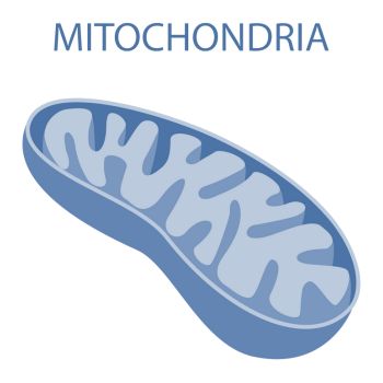
The mitochondria are small intracellular organelles that provide the energy needed by the cell to function properly. The brain and eyes are the most energy-demanding organs. Mitochondria function as batteries that produce more than 90% of the energy in your body’s cells. Mitochondria play a crucial role in powering the organs that require the most energy—especially your heart, liver, muscles, and brain. To give you an idea of their importance: nearly 40% of every heart muscle cell and about a quarter of each liver cell is composed of these tiny energy generators.
Neurons in the brain and throughout the neurological system rely on the energy produced by mitochondria to communicate with one another. When mitochondrial activity is impaired, neurons do not have the energy required to function correctly.
Neurodegenerative diseases gradually disrupt how nerve cells in the brain work, eventually causing these cells to die. Over time, this ongoing loss of brain cell function leads to noticeable declines in memory, movement, or other abilities. In Alzheimer’s Disease, for example, it has been observed that neuronal degeneration, which precedes cell death, is accompanied by impaired mitochondrial activity.
Diseases That Can Result from Mitochondrial Disorders
Mitochondrial disorders can sometimes be genetically driven. The effect of impaired mitochondria can result in:
Muscle Disorders (Myopathies): Weakness, fatigue, and muscle cramps are common, sometimes leading to conditions like mitochondrial myopathy.
Heart Disease: Mitochondrial dysfunction can cause cardiomyopathy, which affects the heart’s ability to pump blood efficiently.
Neurodegenerative Diseases: Conditions like Parkinson’s disease, Alzheimer’s disease, and certain types of dementia have been associated with mitochondrial problems.
Liver Disease: Mitochondrial defects can lead to liver dysfunction or failure, especially in children.
Diabetes: There’s a connection between mitochondrial disorders and diabetes, particularly when it develops at a young age.
Kidney Disease: The kidneys are energy-hungry organs, so mitochondrial issues can sometimes cause kidney problems.
Hearing Loss: Some forms of hearing impairment are related to mitochondrial dysfunction.
Strokes and Seizures: Certain mitochondrial disorders can trigger stroke-like episodes or seizures, even in younger people.
Growth and Developmental Delays: In children, mitochondrial problems can slow growth and cause developmental delays.
Eye Diseases
Mitochondrial Disorders That Can Lead to Eye Disease
The eye relies heavily on energy, making it especially vulnerable to mitochondrial dysfunction. When mitochondria fail to manage oxidative stress or regulate cell survival, a range of problems can arise—particularly in the retina and optic nerve.
Leber Hereditary Optic Neuropathy
LHON is a condition that leads to vision loss by damaging the optic nerve. It typically causes sudden, painless loss of central vision in both eyes, often one after the other over days to months. The disease process begins with swelling of the retinal nerve fibers and eventually leads to the loss of retinal ganglion cells in the optic nerve. 1 2
Dominant Optic Atrophy (DOA) (also known as Kjer Optic Neuropathy)
DOA affects the retinal ganglion cells (RGCs) and nerve fiber layer, leading to reduced visual acuity, color vision deficits, and central scotomas. Visual acuity typically decreases over the first two decades of life to a mean of 20/80 to 20/120.
Pigmentary Retinopathy
This is a group of retinal dystrophies characterized by changes in retinal pigmentation and progressive degeneration of the retina, often leading to visual field constriction and central visual acuity loss. This group of diseases can include: Neuropathy, Ataxia, Retinitis Pigmentosa (NARP) and Maternally Inherited Leigh Syndrome (MILS).
Diabetic Retinopathy
Diabetic Retinopathy is the most common cause of blindness among young adults. This condition develops when high blood sugar damages the blood vessels in the retina, gradually affecting vision and, if left untreated, can eventually lead to blindness. The pathogenesis of diabetic retinopathy involves progressive dysfunction of retinal mitochondria, resulting in the death of retinal capillary cells. 3 Other biomarkers of oxidative stress have been identified that may contribute to diabetic retinopathy.
Age-Related Macular Degeneration
AMD is closely linked to problems with mitochondria, especially in the retinal pigment epithelium (RPE). Research shows that damage to these cellular powerhouses contributes to the onset and worsening of AMD. In addition, exposure to light can make mitochondrial dysfunction worse, further harming retinal cells. 4
Chronic Progressive External Ophthalmoplegia
CPEO causes gradual weakness or paralysis of the muscles that control eye movement. This often leads to drooping eyelids (ptosis) and limited ability to move the eyes. While visual acuity is often preserved, severe cases can lead to secondary complications like corneal disease due to incomplete eyelid closure.
Cataracts
The eye’s lens becomes clouded in this common eye disease. Cataracts can also be a feature in some mitochondrial disorders, such as MELAS and Kearns-Sayre syndrome.
Glaucoma
Glaucoma is the second-leading cause of blindness. It is an optic neuropathy that manifests with optic nerve cupping and atrophy similar to what is observed in primary mitochondrial optic neuropathies. 5 The optic nerve contains a high concentration of mitochondria, which makes it especially vulnerable when these energy producers aren’t functioning well. When mitochondrial respiration is impaired, it can specifically harm retinal ganglion cells (RGCs). In fundus imaging, the red, green, and blue color channels help visualize and assess the health of these cells and the optic nerve, which also plays a role in how we perceive color. 6 Developing trabecular meshwork is thought to have particular sensitivity to oxidative stress-induced damage. 7
A recent study found that people with congenital glaucoma had more potentially harmful mitochondrial DNA mutations compared to those without the condition. Researchers believe these mutations may interfere with mitochondrial function in the trabecular meshwork, a part of the eye involved in fluid drainage. 8
Nutrients That Promote and Support Mitochondrial Health and Function
Nutrients that promote energy (ATP) production, Krebs cycle, and overall mitochondrial support directly and indirectly include: CoQ10 (UBQH), PQQ, acetyl-l-carnitine, Vitamin B6, B12, Riboflavin and Thiamine, Magnesium, NADH, NMN, alpha lipoic acid, resveratrol, n-acetyl-cysteine, turmeric (curcumin), ashwagandha, berberine, taurine, vitamin C, zinc.
A new 2025 study further confirms prior research regarding the cause-and-effect link between faulty mitochondria and the memory loss seen in neurodegenerative diseases. This study suggests that mitochondria could be a powerful new target for treatments. The findings not only shed light on the early drivers of brain cell degeneration but also open possibilities for slowing or even preventing diseases like Alzheimer’s. 9 This study focused on stimulating G proteins found in the mitochondria, essential for the modulation of mitochondrial activity in the brain. Stimulating the G proteins in the brain led to the normalization of both mitochondrial activity and memory performance of dementia mouse models.
Lithium Affects the Mitochondria
A team led by Dr. Bruce A. Yankner at Harvard Medical School discovered that giving mice small doses of lithium orotate—similar to the natural levels found in the brain—could actually reverse disease symptoms and restore brain function.
Lithium is widely prescribed for patients with bipolar disorder, and previous research indicated that it held potential as an Alzheimer’s treatment and an antiaging medication. A 2017 study in Denmark suggested the presence of lithium in drinking water might be associated with a lower incidence of dementia.
This new work is the first to describe the specific roles that lithium plays in the brain, its influence on all of the brain’s major cell types, and the effect that its deficiency later in life has on aging. More research needs to be done.
New Alzheimer’s Study on Lithium
Results of the study by Yankner’s lab and researchers at Boston Children’s Hospital and the Rush Alzheimer’s Disease Center in Chicago also suggest that measuring lithium levels might help doctors screen people for signs of Alzheimer’s years before the first symptoms begin to appear. Yankner said doctors might be able to measure lithium levels in the cerebrospinal fluid or blood, or through brain imaging.
In a healthy brain, lithium maintains the connections and communication lines that allow neurons to talk with one another. The metal also helps form the myelin that coats and insulates the communication lines and helps microglial cells clear cellular debris that can impede brain function.
The depletion of lithium in the brain plays a role in most of the deterioration in several mouse models of Alzheimer’s disease.
Loss of lithium accelerates the development of harmful clumps of the protein amyloid beta and tangles of the protein tau that resemble the structures found in people with Alzheimer’s. Amyloid plaques and tau tangles interfere with how nerve cells communicate with each other in the brain, making it harder for messages to pass from one cell to another.
The plaques, in turn, undermine lithium by trapping it, weakening its ability to help the brain function.
Lithium depletion is involved in other destructive processes of Alzheimer’s: decay of brain synapses, damage to the myelin that protects nerve fibers, and reduced capacity of microglial cells to break down amyloid plaques.
Lithium’s pervasive role comes despite the fact that our brains contain only a small amount of it. After examining more than 500 human brains from Rush and other brain banks, Yankner’s team discovered that the naturally occurring lithium in the brain is 1,000 times less than the lithium provided in medications to treat bipolar disorder.
Yankner and his team found that when they gave otherwise healthy mice a reduced-lithium diet, the mice lost brain synapses and began to lose memory. “We found that when we administered lithium orotate to aging mice [that had] started losing their memory, the lithium orotate actually reverted their memory to the young adult, six-month level,” he said.
Lithium orotate helped the mice reduce production of the amyloid plaques and tau tangles, and allowed the microglial cells to remove the plaques much more effectively.
Yankner said one factor that might help lithium orotate reach clinical trials sooner is the small amount of the treatment needed, which could greatly reduce the risk of harmful side effects, such as kidney dysfunction and thyroid toxicity.
Aside from its potential in treating Alzheimer’s, Yankner said lithium orotate might also have implications for the treatment of Parkinson’s disease, an area his lab is investigating.
Food Sources of Lithium
Grains, vegetables, legumes, and some seafood.