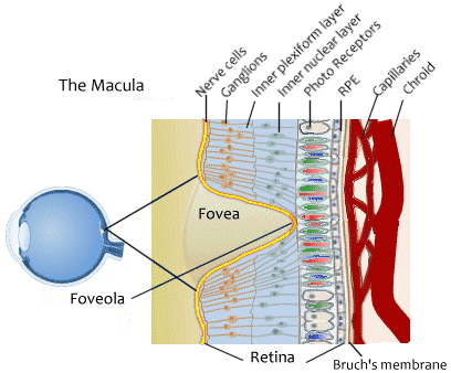
Macular Pigment
How Carotenoids Protect Your Vision
In the 1990s researchers established that the pigments in the retina, composed of carotenoids such as lutein and zeaxanthin, protect the eye from oxidative damage, especially in the macula where higher levels of macular pigment density were found to be inversely related to macular degeneration severity.

In one study1 older subjects with high macular pigment density experienced the same vision sharpness as younger subjects. The researchers found that high macular pigment density was connected to retention of visual acuity, suggesting that macular pigment may retard age-related loss of visual function. People with blue eyes were found to have lower levels of macular pigment making them, especially women, more vulnerable to macular degeneration.
Macular pigment density may be valuable to predict overall eye health, because macular pigment density correlates with preservation of clarity of the lens as well as health of the retina.
Researchers also felt that by improving retina protection through diet (and attending to other risk factors as well) that retinal or retinal pigment epithelial cells that may be damaged but are still viable could recover. It began to appear that patients lose visual acuity before the worst stages of disease but that the worst can be prevented with appropriate diet and lifestyle changes.1
The pigment-related risk factors for macular degeneration are well known and include:
- Light skin color and eye (iris) color
- High exposure to ambient light
- Low levels of dietary yellow pigments (xanthophylls), and low level of xanthophylls in the blood. Lutein, yellow in color, is one of these pigments.
- Cigarette smoking is a strong risk factor and is also associated with lower amounts of macular pigment.
Why macular pigments protect the eye

The pigmented layers of the eye, known as the retinal pigment epithelium (RPE), are not to be confused with the photoreceptor rods and cones of the eye. They act as a color filter through which light passes before it is perceived by the rods and cones. Research in the early 1980s indicated that these pigments consisted of xanthophyll isomers (yellow carotenoids), lutein and zeaxanthin.
Blue light
Yellow carotenoids protect against the damage caused by blue light. Researchers have long understood that plants that gather energy through photosynthesis are protected against oxidative damage by yellow carotenoids. Blue light supports creation of reactive radicals (free radicals), triplet excited states (a form of atom that is readily open to change), superoxide (a form of biologically toxic oxygen), and singlet oxygen in the retina. When retinal pigments are present such creation is slowed or prevented. The yellow pigments that make up macular pigments are effective quenchers of excited triplet states and are reactive toward singlet oxygen and radicals. In that way they actively protect the macula's nerve tissue from the damage.
What else do macular pigments do?
Carotenoids, macular pigments, may also passively shield retinal tissues which are located behind the outer layer of the eye from too much blue light.
Oxidative damage may directly contribute to formation of drusen, the fatty deposits which are a characteristic of macular degeneration.
Researchers have noted that long-term supplementation with lutein may cause a marked increase in the level of pigmentation within the macula.2 Tissues taken from human donor eyes, both controls and those diagnosed with macular degeneration have only 70% as much carotenoids as healthy eyes. The difference is in the entire retina, not only the macula. Therefore researchers concluded that low macular pigment levels were a cause, not a result of degradation of the macular tissue.2
Supplementation with lutein
There is an established association between lutein levels in blood serum and lutein levels in macular tissue. Researchers gave study subjects 30mg of lutein daily over a year period. During that time standardized measurements found that the concentration of lutein in the blood increased ten-fold within the first week and continued to remain high while the subjects continued the supplemental lutein. The process of increase was, after the initial week to 10 days a slow one, amounting to a15%.increase in the pigment level after 72-days of lutein supplementation.2
Lutein and zeaxanthin act synergistically when taken together. For that reason most vision supplements contain these carotenoids along with others for a targeted support approach.
Not all dietary lutein sources are equal
Researchers who explored the ability of carotenoids to increase macular pigment density learned that although leafy greens such as kale supply the largest amounts of lutein and zeaxanthin, these carotenoids provided by orange and red vegetables and fruits are more readily used to support pigment density. 3
Footnotes
1. Schepens Eye Research Institute, An Affiliate of Harvard Medical School, “Improved Nutrition Could Prevent Vision Loss” February 1998.
2. J.T. Landrum, et al, The Macular Pigment: A Possible Role in Protection from Age-Related Macular Degeneration, Advances in Pharmacology, 1997, Volume 38, Pages 537-556.
3. R. Estevez-Santiago, et al., Lutein and zeaxanthin supplied by red/orange foods and fruits are more closely associated with macular pigment optical density than those from green vegetables in Spanish subjects, Nutrition Research, November, 2016.
 info@naturaleyecare.com


 Home
Home


 Vision
 Vision


 Health
 Health
 Research/Services
 Pets
Pets About/Contact


